
Raman Spectroscopy Instruments
 How many cases of prostate cancer occur every year? In 2018, Cancer Journal for Clinicians reported data on 1.276 million new cases of prostate cancer worldwide. What is more, the risk of getting the disease has been rising every year over the past forty years. Therefore, the development of advanced diagnostic methods using sensitive high-tech equipment has become of utmost importance.
In the article "Using the method of «optical biopsy» of prostatic tissue to diagnose prostate cancer," published recently in Molecules, our colleagues investigate the possibility of using optical spectroscopy methods in the differential diagnosis of prostate cancer. Since tumor development brings about structural and biochemical modifications of the tissue, Raman spectroscopy is a promising method of cancer detection and identification.
The scientists studied the prostate glands or their fragments removed during radical prostatectomy on patients with both benign and malignant tumors. Raman spectra were collected from the tissue samples at the excitation radiation wavelengths of 532 and 785 nm. All the samples were also examined by histological methods.
As a result, Raman spectroscopy provided clear evidence of many-fold higher concentrations of cholesterol and its esters (1441 cm-1), as well as fatty acids (1298 cm-1), in tumor cells of adenocarcinoma compared to those of nodular hyperplasia. For example, the level of β-glucose in adenocarcinoma samples was five times higher than in BPH (see Figure 1), which might be related to the lower rate of glucose utilization by tumor cells. The presence of an ensemble of Raman peaks with increased intensity, characteristic of fatty acid, beta-glucose, glutamic acid, and cholesterol, is a fundamental factor in prostate cancer identification.
Thus, the Raman spectroscopy method has been successfully applied in the early diagnosis of prostate cancer, advancing our understanding of the nature of cancerogenesis. Indeed, optical techniques have several advantages compared to traditional imaging methods. Firstly, since optical radiation is non-ionizing, it does not pose any health hazard. Secondly, optical measurements rely on the biochemical and morphological changes in the tissue. Thirdly, owing to the development of fiber-optic probes, Raman spectroscopy can be integrated into endoscopic equipment to enhance the detection of cancer tissue during an operation. In the future, the authors plan to establish correlations between the development of a malignant neoplasm and the changes occurring in the tissue metabolism and biochemical composition of the patient's biological fluids.
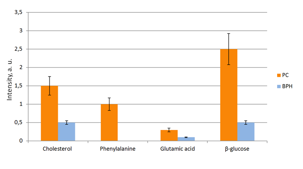 Figure 1. Relative intensities of Raman shifts with error bars denoting several substances identified in the samples of PC (prostate cancer) and BPH (benign prostatic hyperplasia). Level «0» corresponds to the spectrometer noise level. Adopted from here.