
Raman Spectroscopy Instruments
Raman and luminescence spectroscopy is the most advanced modern technology for performing an “optical biopsy” of the tissue. They are used as noninvasive means of diagnosing various diseases and pathological conditions, including tumors of different localizations. Raman spectroscopy relies on the interaction of laser radiation with the tissue. The illuminated sample scatters the light, thereby changing the radiation frequency. Consequently, the frequency shift measured by the spectrometer corresponds to the vibrational and rotational excitations of structural elements of the tissue under investigation. Thus, each substance constituting the tissue is characterized by a set of Raman lines with certain spectral positions and fixed relative intensities. This unique combination of spectral parameters – Raman molecular “fingerprint” – enables the detection of variations in cellular metabolism. Tumor development is accompanied by structural and biochemical modifications of the tissue. Raman spectroscopy registers these changes and makes possible identification of various morphological forms of cancer and concurrent hyperplastic or preneoplastic processes. Raman method proved to be efficient in detecting breast, skin, prostate, cervical, and endometrial cancer.
Optical spectroscopy proved to be an efficient method for the assessment of prostate cancer. The developed analytical PLS-DA method allows to correlate Raman spectra of prostate cancer with validation datasets with 70-80% accuracy. Different wavelengths of laser radiation – 532 nm and 785 nm give different accuracy. Due to the registration of Raman “fingerprints,” the main features of cellular metabolism occurring in the tissue of a malignant prostate tumor were confirmed. Namely: the absence of aerobic glycolysis, over-expression of markers (FASN, SREBP1, stearoyl-CoA desaturase), a strong increase in cholesterol concentration and its esters, as well as fatty acids and glutamic acid. The presence of an ensemble of Raman peaks, inherent in fatty acid, beta-glucose, glutamic acid, and cholesterol, is a fundamental factor for identifying prostate cancer.
Research has shown that Raman spectroscopy differentiates between malignant and healthy breast tissue. From 30 spectra obtained from visually unchanged areas of the mammary gland, 21 spectra corresponded to normal breast tissue; in 8 spectra, the presence of fibrotic areas was determined, and in one spectrum, signs of a pathological process were found. Later, cancer was confirmed (late-stage carcinoma of the ducts) with the help of histological examination. The efficiency of spectral separation of unchanged and metastatic lymph nodes is 85-92%. Thus, Raman spectroscopy has the potential to allow the detailed biochemical assessment of heterogeneous lymph node features, and to contribute towards the development of an optical diagnostic tool for use in a clinical setting.
 Raman Spectral Mapping in the Assessment of Axillary Lymph Nodes in Breast Cancer
J. Smith, C. Kendall, A. Sammon, J. Christie-Brown, N. Stone
Technology in Cancer Research & Treatment

Cervical cancer is the third most common cancer among women worldwide. Most often, the described pathology affects postmenopausal women aged 55 to 62 years. It occurs due to hormonal changes that occur in the female body and provoke various hyperplastic processes in the uterine mucosa. Instant non-invasive express diagnostics using optical spectroscopy makes it possible to identify precancerous conditions.
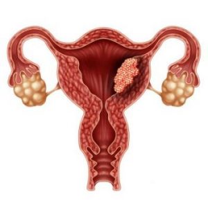
Why does cancer appear? An increased amount of the hormone estrogen produced by the ovaries increases the risk of developing endometrial cancer. If there are too many such hormones in the female body, then the endometrium grows, and its hyperplasia occurs. In this case, the growth of endometrial glands also increases, disrupting cell division and provoking malignant tumors. Another cause of endometrial cancer is mutations in endometrial cells or connective tissue cells, which lead to their atypical growth and the development of a malignant process.
Early diagnostics. Raman spectroscopy is being actively explored as a potential standalone diagnostic tool. Raman represents a unique technology capable of label-free and nondestructively probing endogenous biomolecules, for example, proteins, lipids, carbohydrates, and nucleic acids. Raman spectroscopy has proved to be an efficient method for ex vivo, in vivo, and therapeutic cancer monitoring.