Detection of botulinum neurotoxin type A using SERS

The article «Rapid SERS Detection of Botulinum Neurotoxin Type A» provides an in-depth analysis of a new method for detecting botulinum neurotoxin type A using a SERS technique. The authors highlight the importance of developing rapid and sensitive detection methods for this deadly toxin, which is considered a potential bioterrorism threat.
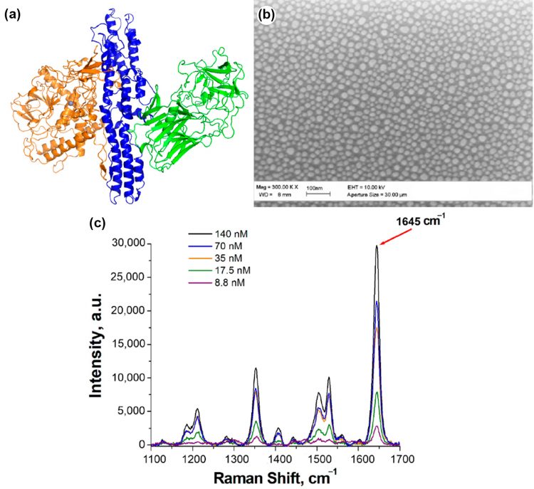
The study presented in the article demonstrates the successful application of SERS for the rapid detection of botulinum neurotoxin type A (see Fig.1 (a)). The authors describe the development of a SERS-based substrate, representing silver nanoislands on Si/SiO2 chip (see Fig.1 (b)), functionalized with an aptamer coupled with a Raman label. The orientation of the label is influenced by the binding of the target, resulting in modifications of the analytical signal (see Fig.1 (c)). These SERS substrates showed high sensitivity and specificity, with a detection limit in the picomolar range.
Furthermore, the article discusses the potential advantages of SERS-based detection, including its rapid analysis time, minimal sample preparation, and the potential for on-site testing. The authors also emphasize the importance of further research and validation of this method to ensure its reliability and practicality in real-world scenarios.
Overall, the scientists provide valuable insights into the development of a promising new approach to the detection of this dangerous toxin. The use of SERS offers a potential solution to the limitations of current detection methods, and its rapid and sensitive nature could significantly improve our ability to respond to potential threats involving botulinum neurotoxin type A. This SERS method serves as an important contribution to the field of toxin detection and has the potential to have a significant impact on public health and safety.
Figure 1. (a) Structure of botulinum neurotoxin serotype A. Receptor-binding domain is shown in green color; the translocating domain is shown in blue color; the catalytic domain is shown in orange color. (b) A SEM image of silver nanoislands on the SERS substrate. (c) SERS spectra of the substrates obtained with different concentrations of Apt 2.22_mod solution. Adopted from here.

